| ID | 112406 |
| Author |
Hisanaga, Satoshi
Tokushima University|Kumamoto University
Miyake, Masato
Tokushima University
Tokushima University Educator and Researcher Directory
KAKEN Search Researchers
Taniuchi, Shusuke
Tokushima University
Morimoto, Masatoshi
Tokushima University
Tokushima University Educator and Researcher Directory
KAKEN Search Researchers
Hirose, Jun
Kumamoto University|Tokushima University
Tokushima University Educator and Researcher Directory
Mizuta, Hiroshi
Kumamoto University
|
| Content Type |
Journal Article
|
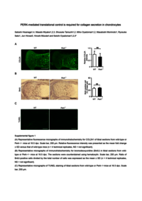
| Description | As chondrocytes are highly secretory and they experience a variety of stresses, physiological unfolded protein response (UPR) signalling is essential for extracellular matrix (ECM) secretion and chondrogenesis. In the three branches of the UPR pathway, PERK governs the translational attenuation and transcriptional upregulation of amino acid and redox metabolism and induction of apoptosis. It was previously demonstrated that a defect of the PERK branch of the UPR signalling pathway causes the accumulation of unfolded proteins, leading to cell death without perturbing endoplasmic reticulum (ER)-to-Golgi transport in pancreatic β cells. However, little is known about the role of PERK in chondrocytes. In this study, we found that PERK signalling is activated in chondrocytes, and inhibition of PERK reduces collagen secretion despite causing excessive collagen synthesis in the ER. Perk−/− mice displayed reduced collagen in articular cartilage but no differences in chondrocyte proliferation or apoptosis compared to the findings in wild-type mice. PERK inhibition increases misfolded protein levels in the ER, which largely hinder ER-to-Golgi transport. These results suggest that the translational control mediated by PERK is a critical determinant of ECM secretion in chondrocytes.
|
| Journal Title |
Scientific Reports
|
| ISSN | 20452322
|
| Publisher | Springer Nature
|
| Volume | 8
|
| Start Page | 773
|
| Published Date | 2018-01-15
|
| Remark | Supplemental figures : srep_8_773_s1.pdf
|
| Rights | © The Author(s) 2018
This article is licensed under a Creative Commons Attribution 4.0 International License, which permits use, sharing, adaptation, distribution and reproduction in any medium or format, as long as you give appropriate credit to the original author(s) and the source, provide a link to the Creative Commons license, and indicate if changes were made. The images or other third party material in this article are included in the article’s Creative Commons license, unless indicated otherwise in a credit line to the material. If material is not included in the article’s Creative Commons license and your intended use is not permitted by statutory regulation or exceeds the permitted use, you will need to obtain permission directly from the copyright holder. To view a copy of this license, visit http://creativecommons.org/licenses/by/4.0/. |
| EDB ID | |
| DOI (Published Version) | |
| URL ( Publisher's Version ) | |
| FullText File | |
| language |
eng
|
| TextVersion |
Publisher
|
| departments |
Institute of Advanced Medical Sciences
Medical Sciences
|