| ID | 106023 |
| Author |
Yasuhara, Yuko
Department of Nursing Management, Institute of Health Biosciences, the University of Tokushima Graduate School
Tokushima University Educator and Researcher Directory
KAKEN Search Researchers
Hirai, Eri
The Major in Nursing, School of Health Sciences, the University of Tokushima
Sakamaki, Sakiko
The Major in Nursing, School of Health Sciences, the University of Tokushima
Tanioka, Tetsuya
Department of Nursing Management, Institute of Health Biosciences, the University of Tokushima Graduate School
Tokushima University Educator and Researcher Directory
KAKEN Search Researchers
Motoki, Kazushi
Department of Clinical Laboratory, Tokushima Prefectural Central Hospital
Takase, Kensaku
Department of Cerebral Surgery, Tokushima Prefectural Central Hospital
Locsin, Rozzano
Department of Nursing, Christine E. Lynn College of Nursing, Florida Atlantic University
Tokushima University Educator and Researcher Directory
KAKEN Search Researchers
Kawanishi, Chiemi
Department of Nursing Art and Science, Institute of Health Biosciences, the University of Tokushima Graduate School
KAKEN Search Researchers
Inui, Tatsuya
Department of Psychiatry, Fujii Hospital
Watari, Chie
Department of Psychiatry, Fujishiro Kensei Hospital
Makiguchi, Kouichi
Department of Psychiatry, Fujishiro Kensei Hospital
|
| Keywords | intramuscular injection
ultrasonography
atypical antipsychotic risperidone long-acting injectable
typical antipsychotic depot intramuscular injection
|
| Content Type |
Journal Article
|
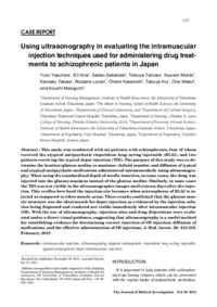
| Description | This study was conducted with six patients with schizophrenia, four of whom received the atypical antipsychotic risperidone long-acting injectable (RLAI), and two patients receiving the typical depot injection (TDI). The purpose of this study was to determine the location (gluteus medius or maximus ; deltoid muscles) and diffusion of typical and atypical antipsychotic medications administered intramuscularly using ultrasonography. When using the standardized depth of needle insertion, in some cases, the drug was injected into the gluteus maximus instead of the gluteus medius. Similarly, in some cases the TDI was not visible in the ultrasonographic images until sixteen days after the injection. This verifies how hard the injection site becomes when microspheres of RLAI is injected as compared to other muscle areas. These results confirmed that the gluteus muscle structure was the ideal muscle for depot injection as evidenced by the injection solution being dispersed and rendered not visible immediately after intramuscular injection (IM). With the use of ultrasonography, injection sites and drug dispersions were evaluated under a direct visual guidance, suggesting that ultrasonography is a useful method for establishing evidence for determining correct insertion of IM injection, diffusion of medications, and the effective administration of IM injections.
|
| Journal Title |
The journal of medical investigation : JMI
|
| ISSN | 13431420
|
| NCID | AA11166929
|
| Volume | 59
|
| Issue | 1-2
|
| Start Page | 213
|
| End Page | 219
|
| Sort Key | 213
|
| Published Date | 2012-02
|
| EDB ID | |
| FullText File | |
| language |
eng
|
| TextVersion |
Publisher
|
| departments |
Medical Sciences
|