| ID | 105999 |
| 著者 |
三田村, 佳典
Department of Ophthalmology, Institute of Health Biosciences, the University of Tokushima Graduate School
徳島大学 教育研究者総覧
KAKEN研究者をさがす
ミタムラ アイザワ, サヤカ
Department of Ophthalmology, Institute of Health Biosciences, the University of Tokushima Graduate School
ナガサワ, トシヒコ
Department of Ophthalmology, Institute of Health Biosciences, the University of Tokushima Graduate School
香留, 崇
Department of Ophthalmology, Institute of Health Biosciences, the University of Tokushima Graduate School
KAKEN研究者をさがす
エグチ, ヒロシ
Department of Ophthalmology, Institute of Health Biosciences, the University of Tokushima Graduate School
KAKEN研究者をさがす
ナイトウ, タケシ
Department of Ophthalmology, Institute of Health Biosciences, the University of Tokushima Graduate School
KAKEN研究者をさがす
|
| キーワード | diagnostic imaging
fundus autofluorescence
optical coherence tomography
retinal photoreceptor cell
retinitis pigmentosa
|
| 資料タイプ |
学術雑誌論文
|
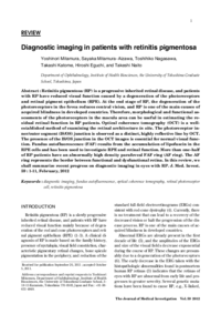
| 抄録 | Retinitis pigmentosa (RP) is a progressive inherited retinal disease, and patients with RP have reduced visual function caused by a degeneration of the photoreceptors and retinal pigment epithelium (RPE). At the end stage of RP, the degeneration of the photoreceptors in the fovea reduces central vision, and RP is one of the main causes of acquired blindness in developed countries. Therefore, morphological and functional assessments of the photoreceptors in the macula area can be useful in estimating the residual retinal function in RP patients. Optical coherence tomography (OCT) is a wellestablished method of examining the retinal architecture in situ. The photoreceptor inner/outer segment (IS/OS) junction is observed as a distinct, highly reflective line by OCT. The presence of the IS/OS junction in the OCT images is essential for normal visual function. Fundus autofluorescence (FAF) results from the accumulation of lipofuscin in the RPE cells and has been used to investigate RPE and retinal function. More than one-half of RP patients have an abnormally high density parafoveal FAF ring (AF ring). The AF ring represents the border between functional and dysfunctional retina. In this review, we shall summarize recent progress on diagnostic imaging in eyes with RP.
|
| 掲載誌名 |
The journal of medical investigation : JMI
|
| ISSN | 13431420
|
| cat書誌ID | AA11166929
|
| 巻 | 59
|
| 号 | 1-2
|
| 開始ページ | 1
|
| 終了ページ | 11
|
| 並び順 | 1
|
| 発行日 | 2012-02
|
| EDB ID | |
| フルテキストファイル | |
| 言語 |
eng
|
| 著者版フラグ |
出版社版
|
| 部局 |
医学系
病院
国際センター
|