| ID | 116982 |
| 著者 |
Yuasa, Masao
University of Tokushima
|
| キーワード | adenocarcinoma in situ
computed tomography
minimally invasive adenocarcinoma
|
| 資料タイプ |
学術雑誌論文
|
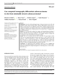
| 抄録 | Background: Given the subtle pathological signs of adenocarcinoma in situ (AIS) and minimally invasive adenocarcinoma (MIA), effective differentiation between the two entities is crucial. However, it is difficult to predict these conditions using preoperative computed tomography (CT) imaging. In this study, we investigated whether histological diagnosis of AIS and MIA using quantitative three-dimensional CT imaging analysis could be predicted.
Methods: We retrospectively analyzed the images and histopathological findings of patients with lung cancer who were diagnosed with AIS or MIA between January 2017 and June 2018. We used Synapse Vincent (v. 4.3) (Fujifilm) software to analyze the CT attenuation values and performed a histogram analysis. Results: There were 22 patients with AIS and 22 with MIA. The ground-glass nodule (GGN) rate was significantly higher in patients with AIS (p < 0.001), whereas the solid volume (p < 0.001) and solid rate (p = 0.001) were significantly higher in those with MIA. The mean (p = 0.002) and maximum (p = 0.025) CT values were significantly higher in patients with MIA. The 25th, 50th, 75th, and 97.5th percentiles (all p < 0.05) for the CT values were significantly higher in patients with MIA. Conclusions: We demonstrated that quantitative analysis of 3D-CT imaging data using software can help distinguish AIS from MIA. These analyses are useful for guiding decision-making in the surgical management of early lung cancer, as well as subsequent follow-up. |
| 掲載誌名 |
Thoracic Cancer
|
| ISSN | 17597714
|
| 出版者 | China Lung Oncology Group|John Wiley & Sons Australia
|
| 巻 | 12
|
| 号 | 7
|
| 開始ページ | 1023
|
| 終了ページ | 1032
|
| 発行日 | 2021-02-17
|
| 権利情報 | This is an open access article under the terms of the Creative Commons Attribution-NonCommercial-NoDerivs License (https://creativecommons.org/licenses/by-nc-nd/4.0/), which permits use and distribution in any medium, provided the original work is properly cited, the use is non-commercial and no modifications or adaptations are made.
|
| EDB ID | |
| 出版社版DOI | |
| 出版社版URL | |
| フルテキストファイル | |
| 言語 |
eng
|
| 著者版フラグ |
出版社版
|
| 部局 |
病院
医学系
|