| ID | 110782 |
| 著者 |
伊東, 進
Department of Digestive and Cardiovascular Medicine, Institute of Health Biosciences, The University of Tokushima Graduate School
徳島大学 教育研究者総覧
六車, 直樹
Department of Digestive and Cardiovascular Medicine, Institute of Health Biosciences, The University of Tokushima Graduate School
KAKEN研究者をさがす
木村, 哲夫
Department of Digestive and Cardiovascular Medicine, Institute of Health Biosciences, The University of Tokushima Graduate School
KAKEN研究者をさがす
矢野, 弘美
Department of Digestive and Cardiovascular Medicine, Institute of Health Biosciences, The University of Tokushima Graduate School
イモト, ヨシタカ
Department of Digestive and Cardiovascular Medicine, Institute of Health Biosciences, The University of Tokushima Graduate School
岡本, 耕一
Department of Digestive and Cardiovascular Medicine, Institute of Health Biosciences, The University of Tokushima Graduate School
徳島大学 教育研究者総覧
KAKEN研究者をさがす
カジ, マサコ
Department of Digestive and Cardiovascular Medicine, Institute of Health Biosciences, The University of Tokushima Graduate School
佐野, 茂樹
Faculty of Pharmaceutical Science, Institute of Health Biosciences, The University of Tokushima Graduate School
徳島大学 教育研究者総覧
KAKEN研究者をさがす
長尾, 善光
Faculty of Pharmaceutical Science, Institute of Health Biosciences, The University of Tokushima Graduate School
徳島大学 教育研究者総覧
KAKEN研究者をさがす
|
| キーワード | infrared fluorescence
infrared fluorescence endoscopy
indocyanin green delivative
endoscopic diagnosis
bioendscopy
|
| 資料タイプ |
学術雑誌論文
|
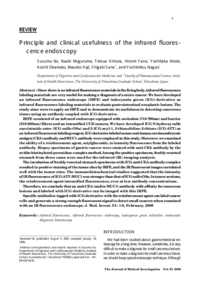
| 抄録 | Since there is no infrared fluorescence materials in the living body, infrared fluorescence labeling materials are very useful for making a diagnosis of a micro cancer. We have developed an infrared fluorescence endoscope (IRFE) and indocyanin green (ICG)-derivative as infrared fluorescence labeling materials to evaluate gastrointestinal neoplastic lesions. The study aims were to apply an IRFE and to demonstrate its usefulness in detecting cancerous tissue using an antibody coupled with ICG-derivative. IRFE consisted of an infrared endoscope equipped with excitation (710-790nm) and barrier (810-920nm) filters and an intensified CCD camera. We have developed ICG N-hydroxy sulfo succinimide ester (ICG-sulfo-OSu) and 3-ICG-acyl-1, 3-thiazolidine-2-thione (ICG-ATT) as an infrared fluorescent-labeling reagent. ICG-derivative-labeled mouse anti-human carcinoembryonic antigen (CEA)antibodyandMUC1 antibody were employed in this study. Moreover, we examined the ability of a reinforcement agent, octylglucoside, to intensity fluorescence from the labeled antibody. Biopsy specimens of gastric cancer were stained with anti-CEA antibody by the avidin-biotinylated peroxidase complex method. Among the positive specimens, freshly resected stomach from three cases were used for the infrared (IR) imaging analysis. The incubation of freshly resected stomach specimens with ICG-anti-CEA antibody-complex resulted in positive staining of the tumor sites by IRFE, and the IR fluorescent images correlated well with the tumor sites. The immunohistochemical studies suggested that the intensity of IR fluorescence of ICG-ATT-MUC1was stronger than that of ICG-sulfo-OSu. In tumor sections, the reinforcement agent intensified fluorescence, ever at low antibody concentrations. Therefore, we conclude that an anti-CEA (and/orMUC1) antibody with affinity for cancerous lesions and labeled with ICG-derivative can be imaged with this IRFE. Specific antibodies tagged with ICG-derivative with the reinforcement agent can label cancer cells and generate a strong enough fluorescent signal to detect small cancers when examined with an IR fluorescence endoscope.
|
| 掲載誌名 |
The journal of medical investigation : JMI
|
| ISSN | 13431420
|
| cat書誌ID | AA11166929
|
| 巻 | 53
|
| 号 | 1-2
|
| 開始ページ | 1
|
| 終了ページ | 8
|
| 並び順 | 1
|
| 発行日 | 2006-02
|
| EDB ID | |
| 出版社版DOI | |
| 出版社版URL | |
| フルテキストファイル | |
| 言語 |
eng
|
| 著者版フラグ |
出版社版
|
| 部局 |
医学系
病院
薬学系
|